Back Of Skull And Neck Anatomy / Osteology Of Head And Neck : They move the head in every direction, pulling the skull and jaw towards the shoulders, spine, and scapula.
byAdmin•
0
Back Of Skull And Neck Anatomy / Osteology Of Head And Neck : They move the head in every direction, pulling the skull and jaw towards the shoulders, spine, and scapula.. The top of the cervical spine connects to the skull, and the bottom connects to the upper back at about shoulder level. The neck is unique in that it supports the weight of your head (10 to 11 pounds) and allows a variety of head/neck movement, such as. The neck is connected to the upper back through a series of seven vertebral segments. The occipital bone is a bone that covers the back of your head; Its clavicular head originates from the medial third of the clavicle, while its sternal head arises from the manubrium of sternum .
Blood vessels of the head and neck. The back muscles stabilize and move the vertebral column, and are grouped according to the lengths and direction of the fascicles. The common cartoid artery extends from the brachiocephalic artery. They move the head in every direction, pulling the skull and jaw towards the shoulders, spine, and scapula. Anatomy the base of the skull is a complex area.
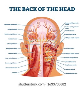
Levator Scapula Muscle And Its Role In Pain And Posture from www.verywellhealth.com The majority of these nerves control the functions of the upper extremities and allow you to feel your arms, shoulder, and back of your head. The neck is connected to the upper back through a series of seven vertebral segments. The large, complex muscles of the neck and back move the head, shoulders, and vertebral column. Think of it like a jigsaw puzzle, all the pieces fit in together and are required to get the full picture as to how it works. The heads come together and ascend diagonally to insert onto the mastoid process of the temporal bone . The muscles of the neck run from the base of the skull to the upper back and work together to bend the head and assist in breathing. These muscles can extend the head, laterally flex it, and rotate it (figure 11.15). The occipital bone surrounds a large opening known as the foramen magnum.
Its clavicular head originates from the medial third of the clavicle, while its sternal head arises from the manubrium of sternum .
A herniated disc between the vertebrae in your neck can cause extreme pain in the base of your skull and back of your neck if the herniated disc presses on a nerve root. In addition, each human skull has a natural bump on the back of the head. The posterior muscles of the neck are primarily concerned with head movements, like extension. The neck is one of the most complex and intricate structures in our body and includes the spinal cord, which sends messages from the brain to the rest of the body. Blood vessels of the head and neck. The occipital bone is a bone that covers the back of your head; In addition, in this region we also find the major cranial and spinal nerves that connect the central nervous system to the organs, skin, and muscles of the head and neck. The skull is a strong, bony capsule that rests on the neck and encloses the brain. It consists of two major parts: The motion of the muscles of the neck are divided into four. Think of it like a jigsaw puzzle, all the pieces fit in together and are required to get the full picture as to how it works. They move the head in every direction, pulling the skull and jaw towards the shoulders, spine, and scapula. The skull can be further subdivided into:
The heads come together and ascend diagonally to insert onto the mastoid process of the temporal bone . See anatomy of the head and neck stock video clips. Anatomy the base of the skull is a complex area. Think of it like a jigsaw puzzle, all the pieces fit in together and are required to get the full picture as to how it works. It extends on each side of the neck and divides at the level of the larynx into two branches:
Neck Nerve High Res Stock Images Shutterstock from image.shutterstock.com The heads come together and ascend diagonally to insert onto the mastoid process of the temporal bone . This bump, called an inion, marks the bottom of the skull where it attaches to the neck muscle. The large, complex muscles of the neck and back move the head, shoulders, and vertebral column. The splenius muscles originate at the midline and run laterally and superiorly to their insertions. The neck muscles, including the sternocleidomastoid and the trapezius, are responsible for the gross motor movement in the muscular system of the head and neck. It consists of two major parts: Anatomy of back of human neck, anatomy of the back and neck, anatomy of the back of the neck, anatomy of the back of the neck muscles, anatomy of the back of your. A pinched nerve in your cervical spine is called cervical radiculopathy.
Its clavicular head originates from the medial third of the clavicle, while its sternal head arises from the manubrium of sternum .
The occipital bone is the only bone in your head that connects with your cervical spine (neck). From the sides and the back of the neck, the splenius capitis inserts onto the head region, and the splenius cervicis extends onto the cervical region. Blood is supplied to parts within the neck, head and brain through branches of the subclavian and common carotid arteries. The skeletal section of the head and neck forms the top part of the axial skeleton and is made up of the skull, hyoid bone, auditory ossicles, and cervical spine. The cervical spine supports the weight and movement of your head and protects the nerves exiting your brain. The back muscles stabilize and move the vertebral column, and are grouped according to the lengths and direction of the fascicles. One reason for shooting neck pain at the base of your skull is a pinched nerve in your upper spine. This example from gray's anatomy shows the cartilages of the throat and the surface anatomy of the neck, with the prominent sternocleidomastoideus which is often thrown into sharp relief when the head is turned or tilted. Neck anatomy nerves picture there are 8 spinal nerves that originate from the cervical spine. In other words, there is a muscle on the forehead (frontalis) and one on the back of the. These muscles can extend the head, laterally flex it, and rotate it (figure 11.15). Ullrich says that the inflammation in the facet joints in the cervical spine between the shoulders and base of the head causes pain at the back of the head. The skull can be further subdivided into:
A herniated disc between the vertebrae in your neck can cause extreme pain in the base of your skull and back of your neck if the herniated disc presses on a nerve root. The head and neck receives the majority of its blood supply through the carotid and vertebral arteries. Each nerve provides sensation to a specific area of the body called a dermatome. Included in this review are the cervica. Cervical spine anatomy (neck) the cervical spine, your neck, is a complex structure making up the first region of the spinal column starting immediately below the skull and ending at the first thoracic vertebra.
Massage For Neck Pain Headaches Suboccipitals from www.painscience.com 10 causes of bumps on the head Cervical spine anatomy video the cervical spine has 7 stacked bones called vertebrae, labeled c1 through c7. In addition, each human skull has a natural bump on the back of the head. Anatomy of back of human neck, anatomy of the back and neck, anatomy of the back of the neck, anatomy of the back of the neck muscles, anatomy of the back of your. Included in this review are the cervica. The majority of these nerves control the functions of the upper extremities and allow you to feel your arms, shoulder, and back of your head. In basic terms, the neck (cervical spine) joins the shoulders and chest to the head. The skull is a strong, bony capsule that rests on the neck and encloses the brain.
It terminates toward the back of the head, behind the ear.
The skull is a strong, bony capsule that rests on the neck and encloses the brain. The skull can be further subdivided into: A pinched nerve in your cervical spine is called cervical radiculopathy. Blood is supplied to parts within the neck, head and brain through branches of the subclavian and common carotid arteries. It involves the upper cervical spine, facet joints, muscles, tendons, ligaments, and nerves. In addition, each human skull has a natural bump on the back of the head. It terminates toward the back of the head, behind the ear. The splenius muscles originate at the midline and run laterally and superiorly to their insertions. The heads come together and ascend diagonally to insert onto the mastoid process of the temporal bone . They move the head in every direction, pulling the skull and jaw towards the shoulders, spine, and scapula. The head and neck receives the majority of its blood supply through the carotid and vertebral arteries. The common cartoid artery extends from the brachiocephalic artery. Each nerve provides sensation to a specific area of the body called a dermatome.
The occipital bone is a bone that covers the back of your head; back of skull anatomy. It involves the upper cervical spine, facet joints, muscles, tendons, ligaments, and nerves.

:max_bytes(150000):strip_icc()/GettyImages-499158129-56a05f075f9b58eba4b0267f.jpg)